
1 min read
Optimizing Imaging Systems for Multiple Procedures, Part 2: Enhanced Patient Experience
This is Part 2 in a 5-part series about optimizing multi-procedure imaging systems. Part 1 offered an overview of the benefits. In Part 2, we’ll...
4 min read
Endovaginal ultrasounds, also known as transvaginal ultrasounds, are a crucial diagnostic tool in gynecology and obstetrics. These ultrasounds provide detailed images of a woman's pelvic organs, including the uterus, ovaries, fallopian tubes, cervix, and surrounding structures. Understanding the procedure, its benefits, and what to expect can help alleviate any concerns and ensure a smooth experience. Here’s what you need to know about endovaginal ultrasounds.
 1. What is an Endovaginal Ultrasound?
An endovaginal ultrasound is a type of pelvic ultrasound where a probe (transducer) is inserted into the vagina to capture detailed images of the pelvic organs. Unlike abdominal ultrasounds, which use a transducer on the skin’s surface, the endovaginal approach allows for closer proximity to the organs being examined, resulting in clearer and more precise images.
Purpose: The primary purpose of an endovaginal ultrasound is to evaluate pelvic pain, abnormal bleeding, infertility issues, and early pregnancy conditions. It is often used to diagnose conditions such as ovarian cysts, fibroids, pelvic inflammatory disease (PID), and ectopic pregnancies.
Procedure: The procedure is typically performed by a sonographer or a gynecologist in a clinical setting. The patient lies on an examination table with their feet in stirrups, similar to a pelvic exam. The transducer, which is covered with a disposable sheath and lubricated, is gently inserted into the vagina. The sonographer then moves the transducer to capture images of the pelvic organs. The entire procedure usually takes about 15 to 30 minutes.
Endovaginal ultrasounds offer several advantages over other imaging modalities, making them an essential tool in women’s healthcare.
High-Resolution Images: The proximity of the transducer to the pelvic organs allows for higher resolution images compared to abdominal ultrasounds. This high level of detail is crucial for accurate diagnosis and monitoring of various conditions.
 Early Detection: Endovaginal ultrasounds can detect early-stage conditions that may not be visible on other types of imaging. This early detection is vital for timely treatment and better outcomes, especially in cases of ectopic pregnancies or early-stage ovarian cancer.
No Radiation: Unlike X-rays or CT scans, ultrasounds do not use ionizing radiation. This makes endovaginal ultrasounds a safer option, especially for pregnant women and those requiring frequent imaging.
Real-Time Imaging: The real-time imaging capability allows for immediate assessment and diagnosis. Physicians can observe the pelvic organs in motion, assess blood flow using Doppler ultrasound, and make prompt decisions about further tests or treatments.
Guidance for Procedures: Endovaginal ultrasounds are often used to guide certain medical procedures, such as egg retrieval for IVF, biopsies, or the placement of intrauterine devices (IUDs). The precise imaging ensures that these procedures are performed accurately and safely.
Proper preparation can help ensure a smooth and successful ultrasound experience. Here are some key points to consider:
Clothing: Wear comfortable clothing that is easy to remove. You may be asked to undress from the waist down and will be provided with a gown or sheet for coverage.
Bladder: Unlike abdominal ultrasounds, which often require a full bladder, endovaginal ultrasounds typically require an empty bladder for better imaging. Follow the specific instructions provided by your healthcare provider.
Medical History: Inform your healthcare provider of any medical conditions, allergies, or current medications. This information helps the provider understand your health status and any potential concerns.
Relaxation: Try to stay relaxed during the procedure. Tension and anxiety can cause discomfort and make the insertion of the transducer more difficult. Deep breathing and focusing on a calm mindset can help ease any discomfort.
Understanding what happens during the procedure can help alleviate any fears or anxieties you may have.
Insertion: The sonographer will gently insert the lubricated transducer into the vagina. You may feel some pressure, but it should not be painful. If you experience significant discomfort, inform the sonographer immediately.
Imaging: The sonographer will move the transducer to obtain different views of the pelvic organs. You may feel slight movements, but these should not cause pain. The sonographer may ask you to adjust your position slightly to get better images.
Communication: Throughout the procedure, the sonographer will explain what they are doing and may ask you about any sensations you feel. Open communication helps ensure your comfort and the accuracy of the procedure.
Post-Procedure: After the procedure, you can resume normal activities immediately. There is no downtime or recovery period required. Your healthcare provider will discuss the results with you and any further steps if necessary.
The results of an endovaginal ultrasound can provide critical insights into your reproductive health.
 Normal Findings: Normal results indicate that the pelvic organs appear healthy with no signs of abnormalities. Your healthcare provider will discuss any routine follow-up care if needed.
Abnormal Findings: Abnormal results may indicate conditions such as ovarian cysts, fibroids, endometriosis, pelvic inflammatory disease, or early pregnancy complications. Depending on the findings, your healthcare provider may recommend further tests, treatments, or referrals to specialists.
Follow-Up: In some cases, a follow-up ultrasound may be necessary to monitor a condition over time or to assess the effectiveness of a treatment. Your healthcare provider will provide guidance on the appropriate follow-up care based on your specific situation.
It’s natural to have questions or concerns about undergoing an endovaginal ultrasound. Here are answers to some common concerns:
Discomfort: While some women may experience mild discomfort during the procedure, it should not be painful. Communicate with the sonographer if you feel significant discomfort, so they can make adjustments to ensure your comfort.
Privacy: Medical professionals conducting the ultrasound are trained to respect your privacy and dignity. You will be draped appropriately, and the procedure will be conducted with sensitivity and professionalism.
Safety: Endovaginal ultrasounds are considered safe and have been used for decades without known risks. The procedure does not involve radiation and has no known adverse effects on the body.
Embarrassment: It’s normal to feel a bit embarrassed about the procedure, but remember that healthcare providers perform these exams regularly and are focused on your health and well-being. Their priority is to make you feel as comfortable as possible.
Endovaginal ultrasounds are a vital diagnostic tool that provides detailed insights into a woman's pelvic health. Understanding the procedure, its benefits, and what to expect can help ease any anxieties and ensure a positive experience. With their high-resolution imaging, early detection capabilities, and safety, endovaginal ultrasounds play a crucial role in women's healthcare. If you have any concerns or questions about the procedure, don't hesitate to discuss them with your healthcare provider. Their goal is to provide you with the best care possible, ensuring your health and peace of mind.
1 min read
This is Part 2 in a 5-part series about optimizing multi-procedure imaging systems. Part 1 offered an overview of the benefits. In Part 2, we’ll...
1 min read
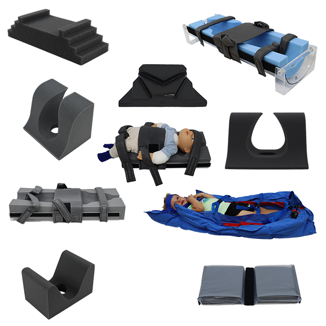
Pediatric imaging presents unique challenges and potential safety risks that require careful consideration and specialized equipment. Unlike adults,...
1 min read
When it comes to radiology and imaging procedures, patient comfort and safety are paramount. One often-overlooked but crucial aspect of this is the...